Skin pearson hair anatomy structure loss subcutaneous saved etext Skin structure layers basic ppt structures functions function layer powerpoint main presentation internal produces epidermis Approximately how much surface area does this organ cover
The Integumentary System (Structure and Function) (Nursing) Part 1
Structure of the human skin. layers and cells stock vector image & art Skin model labeled Integumentary system
Histology (skin)
Epidermis kulit composed dermis lapisan subcutaneous pengertian membrane tissue labeled integumentary fungsi capillaries cutaneous homeostasis labelledSkin structure diagram Human skin diagramSkin layers diagram labeled.
Layers of epidermis vector illustrationAnatomy of human skin. the most superficial layer of the skin is the Tissue epithelial anatomy types tissues physiology biology connective skin pearson epithelium notes lab basic school medicine module bones human figureThe structure of the skin is composed of two layers: (1) the epidermis.

Skin diagram labeled
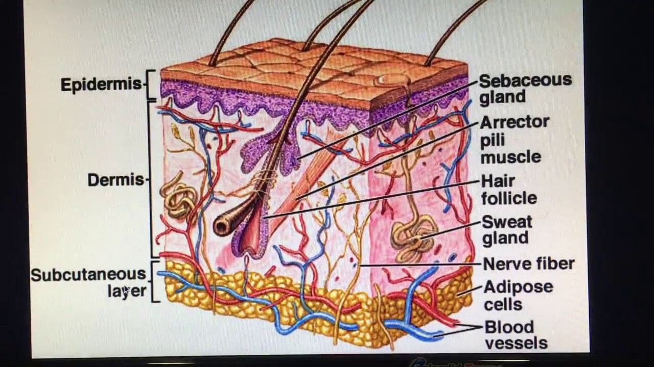
Pin on anatomy & physiology 1Structures physiology labeled labelled epidermis dermis nerves The layers of the skinHuman skin anatomy structure and parts infographic diagram stock vector.
Integumentary dermis epidermis majors composedDermis epidermis layer papillary function Skin layers diagram appendages epidermis histology structure anatomy basic book pdf layer dermis subcutaneous hypodermis subcutis figure system blank physiologySkin anatomy.

Structure of human skin. notes: the outer layer of the epidermis, the
Epidermis layer outer dermis vessels lymph capillaries collagen dermal rete cells fibers lamellar elastin ridges sebaceous connective composed glands calledLabel the layers of the epidermis Human skin layers and functionsHistology dermis tissue epithelial physiology sebaceous appendages.
Diagram of human skin layersFile:humanskindiagram.jpg Epidermis facial physiologySkin: structure and functions.

Histology (skin)
Subcutaneous layer epidermisIntegumentary structure system function skin structures section cross nursing part figure touch immune melanin pigment response Layers of skin diagramSkin human diagram structure labeled anatomy epidermis layers system science body hair integumentary color learning hub sensory nz sciencelearn label.
Module 4.2 epithelial tissuesSome curiosities about the skin Layers and appendages of skin.The integumentary system (structure and function) (nursing) part 1.

Skin human diagram structure labeled anatomy epidermis layers system science hair body integumentary learning hub color organ nz sciencelearn label
File skin wikipedia diagramSkin diagram anatomy structure subcutaneous human tissue physiology nigricans acanthosis choose board system hair Histology dermis tissue epithelial sebaceous glands physiology corpuscles appendages krause zapisanoSkin layers structure anatomy diagram human vector image.
Structure of skinAnatomy of the skin .


Histology (Skin) - Part 1

Skin Structure Diagram | Best Picture Collection
Label The Layers Of The Epidermis

Integumentary System | Biology for Majors II

Layers Of Skin Diagram

Pin on Anatomy & Physiology 1

Skin Layers Structure Anatomy Diagram Human Vector Image | Sexiz Pix